
Comparison of computed tomography and magnetic resonance imaging based target delineation in radiotherapy planning of central nervous system tumors ( High grade gliomas).
DOI:
https://doi.org/10.21276/8wfm0219Abstract
Aim of study : The study was designed to assess and analyze the difference in tumor volumes depicted on computed tomography (CT) and magnetic resonance (MRI) and their effect on the treatment planning.
Materials and methods: twenty -five patients with high grade glioma who had underwent surgical resection and reffered for radiation treatment were taken in the study. CT and MRI imaging were done for all the patients with 1.25 mm thickness. the CT and MRI tumor volumes were delineated and comparision was done between them.
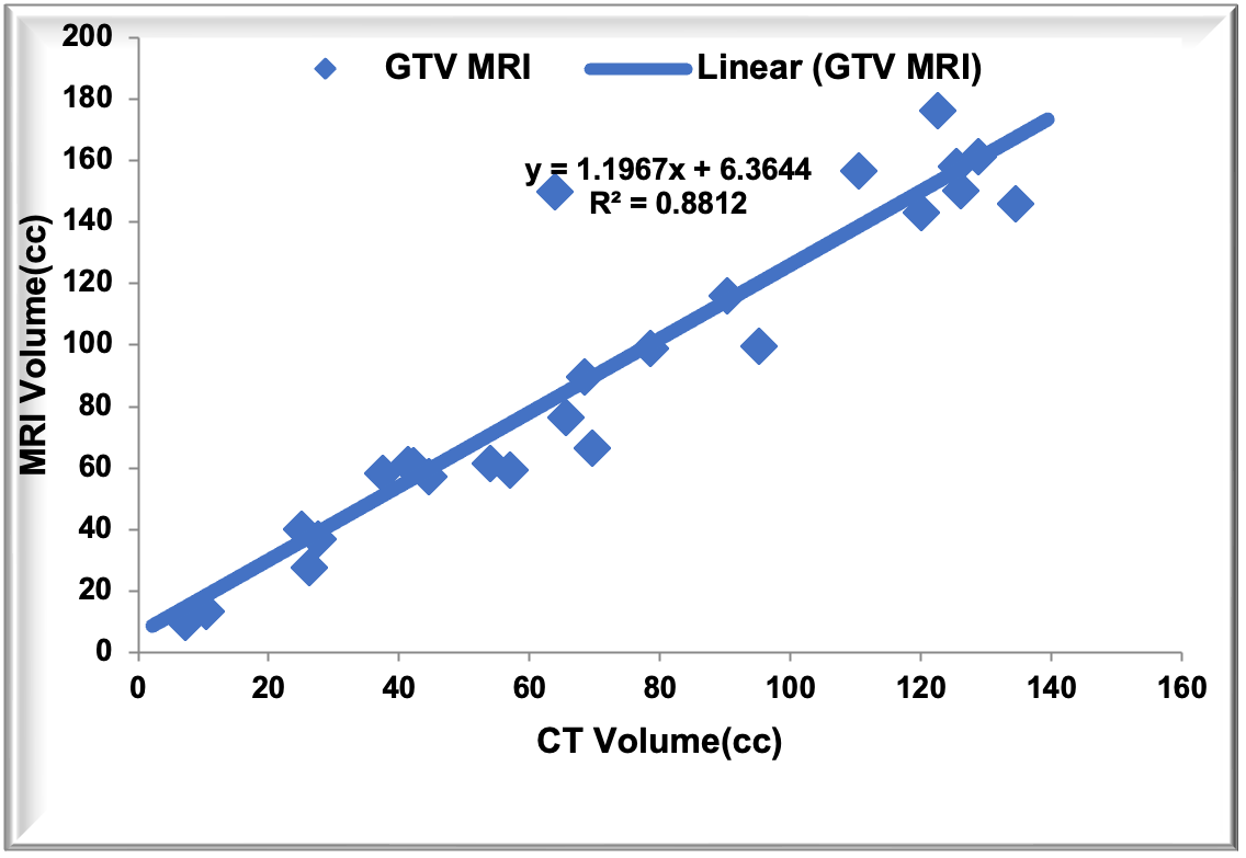
Results: The mean and median of GTV on CT scan were 70.82 and 65.65 respectively. The mean and median of GTV on MRI were 91.11 and 76.54 respectively. A linear relationship between CT and MRI volumes with correaltion coefficient of r=0.93 and MRI shows 1.19 times more volume when comparedwith CT volumes.
Conclusion : The study shows that MRI is an important modality to delineate the brain tumor to avoid the geographical miss and under dosage of the tumor.
key words: high grade glioma, computed tomography, magnetic resonance imaging.
Downloads
References
Kent DL, Haynor DR, Longstreth WT, Larson EB. The clinical efficacy of magnetic resonance imaging in neuroimaging. Ann Intern Med . 1994; 15;120:856-71.
Thornton A.F, Sandler HM, Ten Haken RK, etal. The clinical utility of MRI in 3 -dimensional treatment planning of brain neoplasms. Int J Radiat Oncol Biol Phy 1992; 24:767-75.
Nelson SJ. Multivoxel resonance spectroscopy of brain tumors. Mol Cancer Ther 2003; 2(5) : 497-507.
Websy G, Adamis MK, Edelmann RR. Artifacts in MRI : Description , causes and solutions . In: Edelmann RR, Hessselink JK, Zlatkin MB, editors. Clinical Magnetic Resonance Imaging. Saunders: Philadelphia, Pa; 1996.p.88-144.
Khoo VS, Dearnaley DP,Finnigan DJ, Padhani A, Tanner SF, Leach MO. Magnetic resonance imaging ( MRI) : Considerations and applications in radiotherapy treatment planning. Radiother Oncol 1997; 42:1-15.
Ten Haken RK, Thornton AF Jr, Sandler HM, LaVigne ML. Quint DJ. Fraass BA, etal. A quantitative assessment of the addition of MRI to CT based , 3 D Treatment planning of brain tumors. Radiother Oncol 1992;25:121-33.
Prabhakar R, Haresh KP, Ganesh T, Joshi RC, Julka PK, Rath GK. Comparison of computed tomography and magnetic resonance based target volume in brain tumors. J Cancer Res Ther.2007; volume 3, 121-23.
Downloads
Published
Issue
Section
License
Copyright (c) 2025 karuna singh, NIHARIKA MITTAL, pardeep sharma, sheetal sachdeva (Author)

This work is licensed under a Creative Commons Attribution-NonCommercial 4.0 International License.
Authors are required to sign and submit the completed “Copyright transfer Form” upon acceptance of publication of the paper. This is determined by a publishing agreement between the author and International Archives of Biomedical and Clinical Research. These rights might include the right to publish, communicate and distribute online. Author(s) retain the copyright of their work. International Archives of Biomedical and Clinical Research supports the need for authors to share, disseminate and maximize the impact of their research.